English
 Deutsch
Diagnose Morbus Ormond
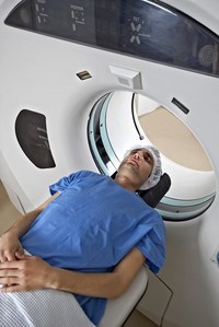
CT and MRI examination
In the event of an onset of the disease, connective tissue in the back abdomen is detected on the basis of a CT or MRI examination.
On the basis of contrast media, a room front in the back of the abdomen becomes visible.
Ultrasound examination
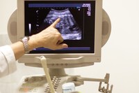
With an ultrasound examination, you can detect a kidney jam that is caused by the compression of one or both ureters. Even during a follow-up examination, ultrasound is looked at, both kidneys work properly and there is no further renal congestion caused by the MO.
If the MO is detected too late, damage to the kidneys can occur in the worst case.
Blood test:
With a boost of the disease, the CHP value is significantly increased.
In the event of a urinary runoff disorder, creatinine and urea levels may be elevated.